|
Locomor problems
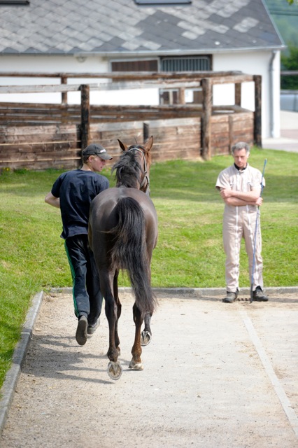
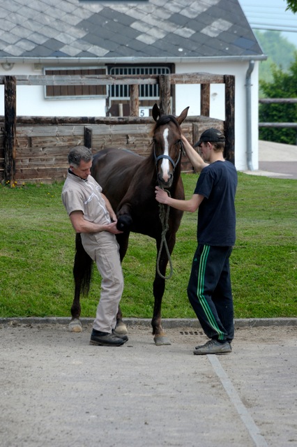
Musculoskeletal problems are the main cause of non-performance in the race horse and in the sport horse. These problems are the main activity of the clinic. One building is dedicated to lameness investigation and imaging : it has 3 consultation rooms, an X-ray room and an ultrasound room. The horses can be observed in straight line and in circle on the hard surface and soft surface.
Radiography
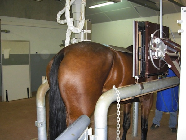
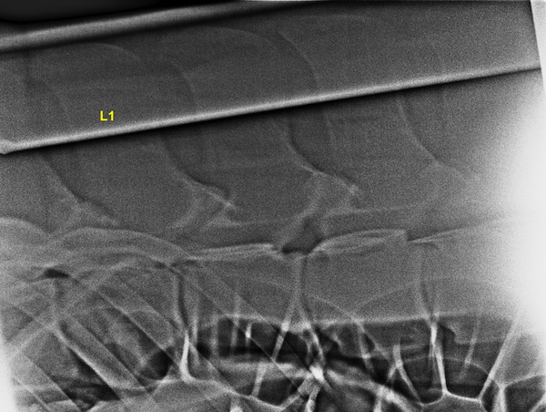
The clinic has a generator mounted on a column that allows radiographic examination of the entire body including back, neck and head. Digital radiography (Fuji Profect, dual-reading) provides high resolution with smaller constant than conventionnal radiography. To maintain the technical facilities, a veterinarian has the title of person competent in radiation protection.
The principle of radiography is based on X-ray attenuation by tissue; when the tissue is more dense, more rays are attenuated and the image is white. X-rays are potentially hazardous to health, protection with aprons and thyroid protectors is essential.
Ultrasonography
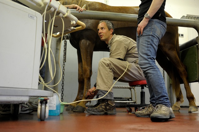
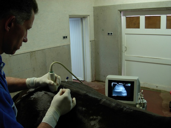
For several years, ultrasound examination became a valuable complement to radiographic examination to examine soft tissues, joints, tendons, ligaments, muscles and regions such as deep cervical joints, lumbar and sacroiliac regions.
The clinic is equipped with an ultrasound system (Philips) with three probes (tendon, back, and pastern) for the diagnosis and to perform ultrasound-guided infiltration. Two additional systems with rectal probes allow the examination of sacroiliac and lumbosacral regions. Another system is present in the surgery building to diagnose some specific diseases.
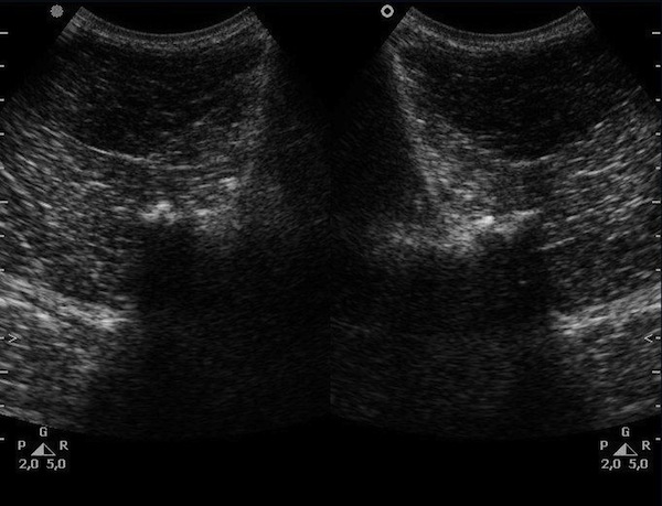
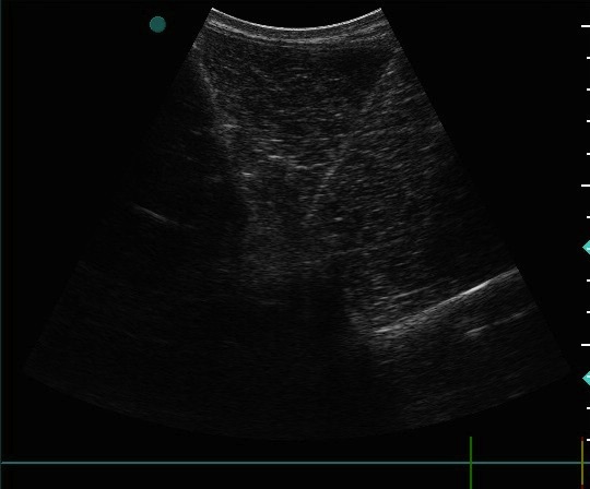
The principle is based on the emission of a ultrasound wave by the probe which is transmitted in the tissues. The echoes pass through the water but are reflected by the high-density tissues and air. The echoes are received by the probe and converted into an image that shows the position and density of tissues.
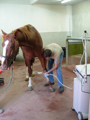
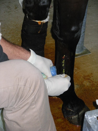
Infiltration
The clinic provides all routine injections needed to treat different problems. For deep infiltration (neck, back, pelvis, shoulder), injections are performed under ultrasound guidance to confirm the position of the needle in the right place.
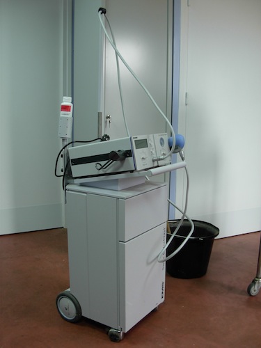
Shockwave
The shock wave therapy is used to treat certain diseases including ligamentous insertions such as insertions of the suspensory ligament (body and branches). Each treatment includes several sessions with a number of precise blows.
The shock waves are emitted directly onto the affected area. This causes an increase in blood flow, activation of osteogenic factors and a direct analgesic effect (pain suppression).
IRAP
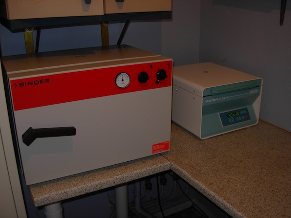
IRAP (Interleukin-1 Receptor Antagonist Protein) is used for several years for the treatment of osteoarthritis. The main principle of treatment is to block the effect of interleukin-1 (molecule generating inflammation) using the serum of the horse.
For this, 50 ml of blood are directed into a syringe containing glass beads. The blood is incubated for 24 hours to form IRAP and is then centrifuged to pack the samples. Conventional treatment includes a series of multiple injections. The IRAP is no doping and has no side effects on cartilage.
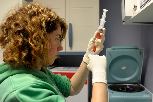 
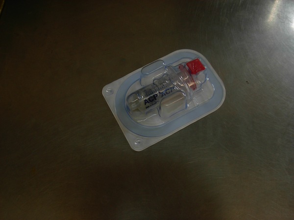
PRP
PRP (Platelet Rich Plasma) is used for treatment of tendonitis and desmitis. The main principle is to concentrate platelets from the blood which contains a number of growth factors. These factors allow formation of new blood vessels, formation of new connective tissue and regeneration.
For this, 15 ml of blood are centrifuged to isolate the fraction containing platelets. Injection can be performed on standing horses or under general anesthesia depending on the location of the lesion. Bone marrow or bone marrow aspirate concentrate can be injected at the same time to bring stem cells on the healing tissue.
Stem cells
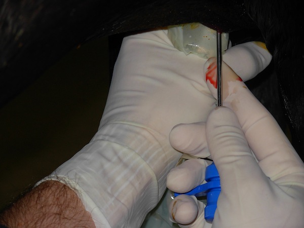
Stem cells allow to improve healing. They are used to treat tendonitis or some joint problems which do not respond to usual treatments.
For this, 20 ml of bone marrow are isolated from the sternum or pelvis. Bone marrow is sent to laboratories where stem cells are isolated. Two or three weeks are needed for that. The sample is then injected in the tendon under ultrasonographic guidance or in the joint.
|
 Lameness investigation